Training

Announcing a new short course series in utilizing the advanced instrumentation and expertise available in the Imaging and Analysis Center
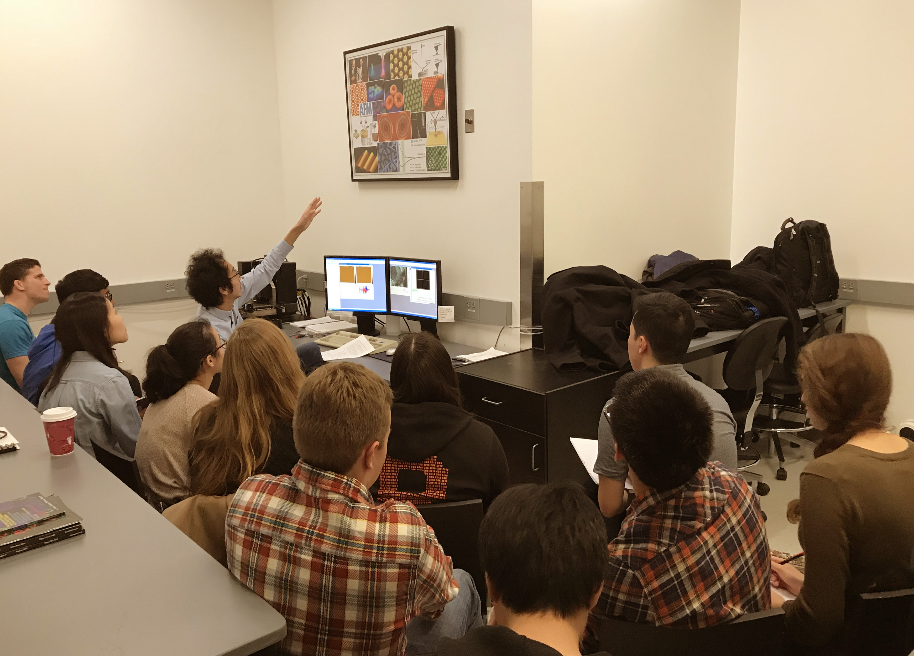
The Imaging and Analysis Center (IAC) at the Princeton Institute for the Science and Technology of Materials, Princeton University, welcomes the opportunity to facilitate broad usage of its state-of-the-art instrumentation and expertise in materials research and education. As a crucial component of this effort, the IAC is initiating a new program to further promote education in experimental methods of imaging and analysis. We will provide a series of group training courses for future IAC users to the Princeton community. The training courses involve direct experimental demonstrations and hands-on instruction in the use of all IAC equipments ranging from basic sample preparation device to high-end electron microscopes. These 3-6 hour courses are offered regularly and frequently, so that users can receive basic training when they anticipate an imminent need for the technique. The training courses thus complement PRISM's "for-credit" course, MSE 505 (Characterization of Materials) which covers the principles and applications of all these techniques in depth. The topics are listed below.(Click on the individual topic to get more information and the training schedule)

Safety is a top priority at Princeton University. Please take time to review the Environmental Health and Safety website.


To use IAC equipment and sign up for training courses... Register an email address here
Need assistance with IAC equipment? Click here!
Check out our current Instrument Rates
All training courses will begin outside ACEE 032.

Basic sample preparation for microscopy

1a. Sample preparation for SEM, XRD, Microprobe and FIB
1b. TEM sample preparation for soft materials (by ultramicrotomy)
1c. TEM sample preparation for hard materials (by ion milling)
1d. TEM sample preparation for biological or soft materials (by staining and/or resin embedding)
1e. TEM sample preparation for biological or soft materials (by plunge-freezing)

Basic operation of various imaging and analysis instruments

2a. Operation of an x-ray diffractometer (Rigaku Miniflex XRD)
2b. Operation of a scanning electron microscope (XL30 FEG-SEM)
2c. Operation of Transmission Electron Microscope (Philips CM200 TEM)
2d. Operation of an atomic force microscope (Bruker Multimode AFM)
2e. Operation of an Ellipsometer (M-2000)
2f. Operation of a Raman Spectrometer (Horiba)
2g. Operation of X-ray Photoelectron Spectrometer (Thermo K-alpha XPS/UPS)
2h. Operation of a Leica DCM3D Confocal Micro-optical System
2i. Operation of a Photoluminescence Spectrometer (Edinburgh Instruments, FLS980)
2j. Operation of a UV-Vis Spectrometer (Agilent Technologies, Cary 5000)
2k. Operation of a Rheometer (Anton Paar MCR501)
2l. Operation of Thermo Analysis Equipment (DSC, DMA)
2m. Operation of Thermo Analysis Equipment (TGA-GC/MS)
2n. Operation of a Thermo FTIR Imaging Microscope (Nicolet iN10 MX)

Advanced operation of various imaging and analysis instruments

3a. Operation of Energy Dispersed X-ray Spectroscopy for Microanalysis (Oxford AZtech EDS operated with XL30 FEG-SEM)1
3b. Operation of an environmental scanning electron microscope (Quanta 200 FE-ESEM)1
3c. Operation of High-resolution, Low-voltage Scanning Electron Microscope (FEI Verios 460 XHR SEM)1
3d. Operation of a high-resolution x-ray diffractometer (Bruker D8 Discover)2
3e. Operation of 3D X-ray Microscope (Zeiss Xradias Versa 520)2
3f. Operation of Wave Dispersed X-ray Spectroscopy and Electron BackScattering Diffraction (WDS and EBSD operated with Quanta FE-ESEM)3
3g. Operation of Focused Ion Beam System (FEI Helios G3 DualBeam FIB/SEM)4
3h. Operation of Dimension Atomic Force Microscope (Bruker Dimension NanoMan AFM)5
3i. Operation of Dimension Atomic Force Microscope (Bruker Dimension ICON3 AFM)5
3j. Operation of High-resolution Scanning Transmission Electron Microscope (Talos F200X S/TEM with SuperX-EDS)6
3k. Operation of Ultra High-resolution Double Cs-corrected Scanning Transmission Electron Microscope (Titan Themis 80-300 Cubed S/TEM)7
3l. Operation of a Twin-Motor Rheometer (Anton Paar MCR702)8

1. Attendants are required to have previous training and practice in use of the XL30 SEM (2b).
2. Attendants are required to have previous training and practice in use of the Rigaku Miniflex XRD (2a).
3. Attendants are required to have previous training and practice in use of the Quanta 200 FE-ESEM (3b).
4. Attendants are required to have previous training and practice in use of the Verios 460 XHR SEM (3c).
5. Attendants are required to have previous training and practice in use of the Bruker Multimode AFM (2d).
6. Attendants are required to have previous training and practice in use of the CM 200 TEM (2c).
7. Attendants are required to have previous training and practice in use of the Talos F200X S/TEM (3j).
8. Attendants are required to have previous training and practice in use of the Anton Paar MCR501 (2k)..
* courses coming soon